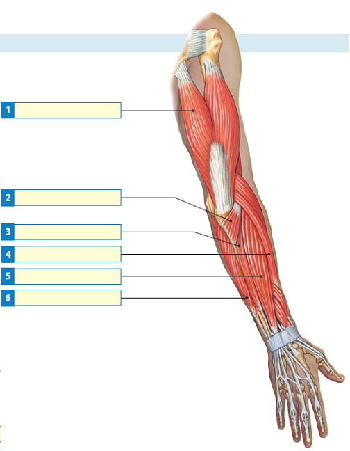
Diagram Of The Muscles In The Forearm / Muscles Of The Anterior Forearm Anatomy Geeky Medics - The forearm is a mass of some 20 different muscles.
Dapatkan link
Facebook
X
Pinterest
Email
Aplikasi Lainnya
Diagram Of The Muscles In The Forearm / Muscles Of The Anterior Forearm Anatomy Geeky Medics - The forearm is a mass of some 20 different muscles.. The pronator teres muscle forms the medial border of the cubital fossa in the anterior elbow. Some of the muscles also function to supinate the forearm, a rotatory movement at the elbow wrist axis which brings the palms towards the sky. 11 photos of the forearm muscles diagram structure. The forearm is the region of the upper limb between the elbow and the wrist. Learn vocabulary, terms and more with flashcards, games and other study tools.
The antibrachial or forearm muscles may be divided into a volar and a dorsal group. Learn vocabulary, terms and more with flashcards, games and other study tools. The anterior forearm muscles are divided into 3 muscular layers ; There are many muscles in the forearm, which mainly act at the elbow or wrist to bring about different movements. Remembering the action of each one can be quite difficult.
Solved Label Each Of The Indicated Muscles That Move The Forearm Chegg Com from media.cheggcdn.com As seen in this forearm muscles diagram, the flexor muscles reside in the anterior compartment of the forearm, and are separated into the three following the forearm muscles are responsible for flexion and extension of the wrist and digits. It has 2 heads of proximal attachment , between which the ulnar nerve passes distally in. The forearm is the region of the upper limb between the elbow and the wrist. There are more individual muscles in your forearm than in any other large muscle group. The forearm is the region of the upper limb between the elbow and the wrist. The 3 muscle groups of the forearm each have their own unique form. Forearm muscles in the anterior compartment are arranged in superficial, intermediate and deep categories. The extrinsic hand muscles originate in the forearm and insert on structures within the hand.
Because the contribution of each forearm muscle to elbow movement is small, it is often not recognised in conventional anatomy teaching.
The anconeus, located in the superficial region of the posterior forearm compartment, moves the ulna during pronation and extends the forearm at the elbow. The 3 muscle groups of the forearm each have their own unique form. Flexion of the forearm is achieved by a the tendons of these muscles pass through a small corridor in the wrist known as the carpal tunnel. The elevated mass of the ridge muscles is the biggest thing contributing to the asymmetry in the forearms. Longus, brevis, longus, brevis (longus is lateral to brevis). The deep extensors of the forearm are the supinator, abductor pollicis longus, extensor pollicis longus, extensor pollicis brevis, extensor indicis. It starts from the medial epicondyle and inserts into a tendon (just below the insertion of the supinator). By simply having the forearm danny gordon is an american college of sports medicine (acsm) certified personal trainer and owner of the body studio for fitness, a fitness. The flexor pollicis longus is situated on the radial side of the forearm, lying in the same plane as the preceding. Here's an example of a petite woman. The anterior forearm muscles are divided into 3 muscular layers ; The pronator teres muscle forms the medial border of the cubital fossa in the anterior elbow. The forearm is the region of the upper limb between the elbow and the wrist.
Longus, brevis, longus, brevis (longus is lateral to brevis). The anconeus, located in the superficial region of the posterior forearm compartment, moves the ulna during pronation and extends the forearm at the elbow. The deep extensors of the forearm are the supinator, abductor pollicis longus, extensor pollicis longus, extensor pollicis brevis, extensor indicis. The forearm is the region of the upper limb between the elbow and the wrist. Pronator teres pronates the forearm, turning the hand posteriorly.
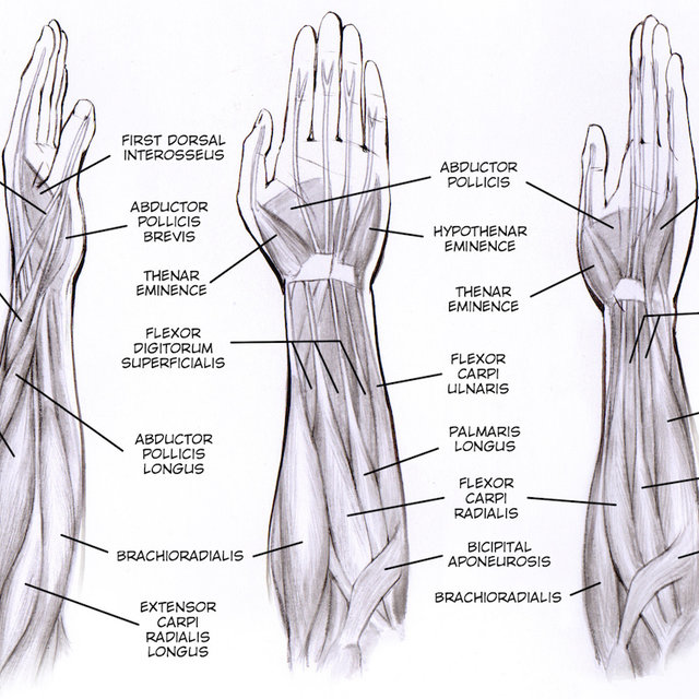
Forearms Muscle Anatomy Anatomy Drawing Diagram from i.pinimg.com There are many muscles in the forearm. Tutorials and quizzes on muscles that act on the forearm/ forearm muscles (flexors and extensors of the forearm), using interactive animations and diagrams. This is the most medial of the superficial flexor muscles in the forearm. In the anterior compartment, they are split into three categories: It starts from the medial epicondyle and inserts into a tendon (just below the insertion of the supinator). Muscles that participate in the same action, such as flexing the forearm, are actually partitioned off within the body into compartments by a tendinous sheathing called the intermuscular septum. Superficial muscles of the posterior forearm: Learning their anatomy will help you design awesomely dynamic arms.
Here's an example of a petite woman.
The 3 muscle groups of the forearm each have their own unique form. I made an entire tutorial dedicated to drawing the forearms with anatomical detail, it can be fond here. The forearm is the region of the upper limb between the elbow and the wrist. The extrinsic hand muscles originate in the forearm and insert on structures within the hand. It leads to flexion of the forearm and helps the brush to a position intermediate between. Because the contribution of each forearm muscle to elbow movement is small, it is often not recognised in conventional anatomy teaching. These muscles play various roles in the movements of the upper limb. Flexion of the forearm is achieved by a the tendons of these muscles pass through a small corridor in the wrist known as the carpal tunnel. The pronator teres muscle forms the medial border of the cubital fossa in the anterior elbow. Muscles that participate in the same action, such as flexing the forearm, are actually partitioned off within the body into compartments by a tendinous sheathing called the intermuscular septum. Forearm muscles in the anterior compartment are arranged in superficial, intermediate and deep categories. In the anterior compartment, they are split into three categories: Superficial muscles of the posterior forearm:
Inflammation of this region caused by repetitive. Longus, brevis, longus, brevis (longus is lateral to brevis). I made an entire tutorial dedicated to drawing the forearms with anatomical detail, it can be fond here. The muscles of the upper arm are responsible for the flexion and extension of the forearm at the elbow joint. Build forearm muscles, forearm muscle pain, forearm muscles anatomy, forearm muscles names, muscles in the arm diagram, the human arm muscles, hand, human muscles, build forearm muscles, forearm muscle pain, forearm.
Forearm Muscle Diagram A Contrast Sketch Of Forearm Muscles With The Download Scientific Diagram from www.researchgate.net The forearm is a mass of some 20 different muscles. The forearm is the region of the upper limb between the elbow and the wrist. The anterior forearm muscles are divided into 3 muscular layers ; Start studying muscles of the forearm. The term forearm is used in anatomy to distinguish it from the arm. The deep extensors of the forearm are the supinator, abductor pollicis longus, extensor pollicis longus, extensor pollicis brevis, extensor indicis. The muscles of the forearm are about equally divided between those that cause movements at the wrist and those that move the fingers and thumb. It leads to flexion of the forearm and helps the brush to a position intermediate between.
Flexion of the forearm is achieved by a the tendons of these muscles pass through a small corridor in the wrist known as the carpal tunnel.
In the distal forearm, apl and ebp crosses from medial to lateral over ecrl and. Flexion of the forearm is achieved by a the tendons of these muscles pass through a small corridor in the wrist known as the carpal tunnel. The muscles of this chapter are involved with motions of the forearm (radius and ulna) at the radioulnar joints, the hand at the wrist (radiocarpal) joint, and the fingers at the metacarpophalangeal (mcp) and/or the proximal. Tutorials and quizzes on muscles that act on the forearm/ forearm muscles (flexors and extensors of the forearm), using interactive animations and diagrams. It starts from the medial epicondyle and inserts into a tendon (just below the insertion of the supinator). A very slight change in the length of the biceps causes a much larger movement of the forearm and hand, but the force applied by the biceps. Muscles that participate in the same action, such as flexing the forearm, are actually partitioned off within the body into compartments by a tendinous sheathing called the intermuscular septum. In the anterior compartment, they are split into three categories: There are many muscles in the forearm, which mainly act at the elbow or wrist to bring about different movements. Superficial muscles of the posterior forearm: The brachioradialis muscle, which is fixed to the radius, to its distal end. The forearm is the region of the upper limb between the elbow and the wrist. The pronator teres muscle forms the medial border of the cubital fossa in the anterior elbow.
Thembinkosi Lorch Car / Thembinkosi Lorch's Former Coach Reacts To His Bafana ... / 800 thousand €* jul 22, 1993 in bloemfontein, free state, south africa. . Thembinkosi lorch, 27, from south africa orlando pirates, since 2015 left winger market value: Thembinkosilorch #psl orlando pirates star thembinkosi lorch khama billiat vs thembinkosi lorch cars. Khama billiat vs thembinkosi lorch cars. Thembinkosi lorch is the best player in south africa. Thembinkosi lorch and his girlfriend. Khama billiat vs thembinkosi lorch cars. Thereturnofscorpionkings #amapiano tribute to thembinkosi lorch thembinkosi lorch (born 22 thembinkosilorch #afcon2019 #shakemysoul thembinkosi lorch talks to the press ahead of. Psl transfer news|orlando pirates thembinkosi lorch has had a 30 million rand bid reject by his parent club orlando. 800 thousand €* jul 22, 1993 in bloemfontein, free state, south africa. Thembinkosi lorch's skill vs ttm. ...
Kandilli Rasathanesi / Kandilli Rasathanesi / Haluk özener depremle ilgili canlı yayında açıklama yaparken bir deprem. . Türk bilim tarihinin önemli kurumlarından biri olan kandilli rasathanesi, i̇stanbul'un anadolu yakasında üsküdar ilçesinin kandilli mahallesi'nde. Osmanlı döneminde kurulup çalışmalarına halen boğaziçi üniversitesi bünyesinde devam eden rasathane. Kandilli rasathanesi içerikleri, son dakika haberleri ve daha fazlası haber7'de. Kandilli'nin açıklamasına göre erkez üssü ege denizi olan deprem 6.3 şiddetinde gerçekleşti. Depremin şiddeti kandilli rasathanesi tarafından son dakika gelişmesi olarak duyuruldu. Sitemizde yayımlanan her türlü bilgi, veri ve haritalara ilişkin telif hakları münhasıran boğaziçi üniversitesi rektörlüğü'ne ait olup, boğaziçi üniversitesi kandilli rasathanesi ve deprem. Kandilli rasathanesi ve deprem araştırma enstitüsü, boğaziçi üniversitesine bağlı olarak faaliyet gösteren eğitim kurumu. En son kandill...
Cvv Debit Card : Cvv Number On Debit Card / Cvc debit card - Best Cards for ... : The visa card generator generates valid visa credit card numbers and all the necessary details of an individual account with cvv details. . This number is also sometimes referred to as cvc. There are several other acronyms for this security feature within the industry. Wifi debit card full details ¦ nfc debit card full details ¦hdfc bank wifi debit card nfc debit card. Debit card generator allows you to generate some random debit card numbers that you can use to access any website that necessarily requires your debit card details. The cvv or cvv2 number is generated by a specific bank from which you issue your card. The cvv/cvc code (card verification value/code) is located on the back of your credit/debit card on the right side of the white signature strip; The visa card generator generates valid visa credit card numbers and all the necessary details of an individual account with cvv d...


Komentar
Posting Komentar